Research
Overview
The Clark Lab seeks to understand how cells produce functional proteins at the proper levels, including how the cellular environment supports folding of proteins to their functional (“native”) structures. This work is important because the dysregulation of protein production and/or protein folding underlies a wide variety of human diseases, yet our understanding of the cellular mechanisms that regulate gene expression and protein folding is still rudimentary at best. Although powerful new machine learning approaches like AlphaFold have enabled transformative breakthroughs in protein structure prediction, the experimental datasets that underpin the success of these approaches were not designed to support accurate prediction of the effects of mutations on protein stability and misfolding propensity. These are the knowledge gaps that our lab seeks to fill.
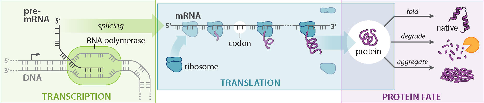
All projects in our lab connect to the key concept that, in the cell, protein folding begins vectorially (meaning, from one end of the protein to the other) as proteins are synthesized by the ribosome and/or translocated across a membrane. In contrast, most of our current understanding of protein folding mechanisms is derived from diluting full length proteins out of a chemical denaturant. Dilution from denaturant enables folding to begin by interactions formed between any two portions of the protein, rather than providing a “head start” to the first portion of the protein to appear. Protein folding initiated during vectorial appearance is thus a fundamentally different starting point than folding initiated by dilution from denaturant and represents the environment under which protein sequences were selected by evolution to fold correctly. Crucially, our lab pioneered new experimental approaches that demonstrated that initiating folding during vectorial appearance can significantly enhance the success of protein folding to the native structure, by suppressing misfolding and aggregation. Approaches developed in our lab are now broadly used by others to study the conformations of co-translational protein folding intermediates.
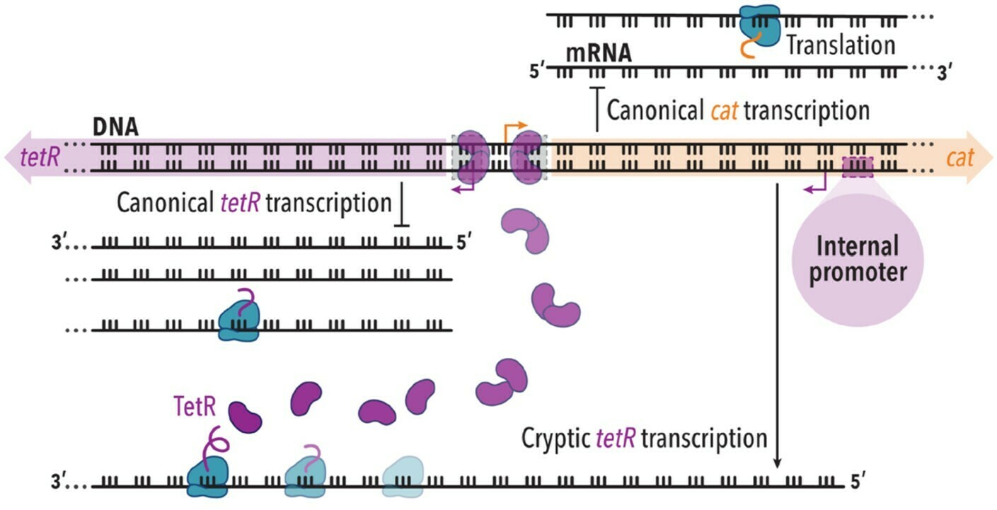
Our studies of co-translational protein folding have sparked a longstanding interest in the impact of synonymous codon usage on cell function. Substitutions between synonymous codons were long thought to be “silent” because these mutations preserve the amino acid sequence of a protein. Indeed, to this day, such mutations are often disregarded, even when associated with disease. However, synonymous substitutions can alter the rate of protein synthesis and therefore the rate at which a protein can interact with other portions of itself. Our lab has pioneered the development of new experimental and computational methods to measure the impact of synonymous codon substitutions. Using these methods, we discovered that synonymous codon substitutions can significantly perturb protein folding mechanisms, including altering whether a protein will fold to its native structure or not. These perturbations can significantly decrease cell fitness. As part of this work, we have also identified novel mechanisms by which synonymous codon substitutions can affect other aspects of gene expression, including transcriptional regulation of neighboring genes.
Our lab pursues multiple projects simultaneously, typically one project per person, but with a lot of synergy between the approaches we use. We rely heavily on experimental biophysical and biochemical approaches, including fluorescence spectroscopy, but we have a strong history of incorporating methods from diverse fields — or developing novel methods when necessary — to answer our research questions. We use the bacterium E. coli as an experimental system to enable deep exploration of fundamental questions regarding the impact of the cellular environment on protein folding, but we routinely analyze human and other eukaryotic genomes to test broad hypotheses regarding the generality of our results. To facilitate our multidisciplinary approach, we collaborate broadly and deeply with experts in diverse fields, including statistics, computer science, genetics, microbiology, structural biology, molecular dynamics, eukaryotic cell biology, analytical chemistry, and graphic design.
Current project areas
Impact of co-translational folding on protein homeostasis
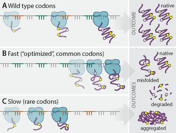
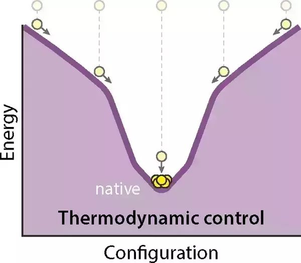
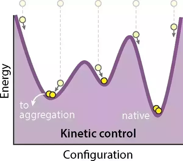
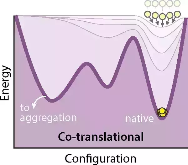
As each protein is synthesized by the ribosome, the nascent polypeptide chain emerges from the ribosome exit tunnel from N- to C-terminus and can begin to fold into its functional three-dimensional structure. The rate of protein synthesis — specifically, the rate of translation elongation — can be slower than the folding rates for some protein secondary and tertiary structures. This means that the amino acid residues near the N-terminus of the nascent polypeptide chain can begin to form stabilizing interactions with other N-terminal residues during translation. This process is called co-translational folding. Co-translational folding can alter the way in which a protein is exposed to its energy landscape. For example, our lab has shown that co-translational folding can prevent diverse proteins from populating unproductive, misfolded conformations on the energy landscape for folding (Evans et al. (2008) “Cotranslational folding promotes beta-helix formation and avoids aggregation in vivo” and Ugrinov & Clark (2010) “Cotranslational folding increases GFP folding yield”). Co-translational folding can also modulate whether a nascent protein will interact co-translationally within other molecules in the cell. In bacteria, the folding of membrane-bound proteins also occurs co-translationally, often in association with the Sec translocon, which facilitates the insertion of proteins into the membrane as they fold. We study diverse aspects of co-translational protein folding, including co-translational assembly of oligomeric proteins and the impact of the rate of protein appearance and folding direction on the success of protein folding. For more information on co-translational protein folding, see the review articles on “The Effects of Codon Usage on Protein Structure and Folding” and “Protein folding success depends on the direction and speed of polypeptide chain appearance.”
Impact of synonymous codon usage on protein homeostasis
In the genetic code, the same amino acid residue can be encoded by more than one tri-nucleotide codon. The set of codons that encoding the same amino acid are termed as synonymous codons. Traditionally considered “silent,” over the past decade our lab and others have conclusively shown that synonymous codon substitutions can significantly influence diverse aspects of protein production, including transcription, mRNA splicing, the efficiency of translation initiation, the accuracy of translation, and translation elongation rate. Our lab published the first study (Sander et al. (2014) “Expanding Anfinsen’s principle: contributions of synonymous codon selection to rational protein design”) demonstrating that synonymous codon substitutions can alter protein folding outcome in a predicable way in vivo. We subsequently showed that synonymous mutations can disrupt co-translational protein folding (Walsh et al. (2020) “Synonymous codon substitutions perturb cotranslational protein folding in vivo and impair cell fitness”), leading to increased protein degradation and reduced cell fitness. Most recently, we discovered that synonymous mutations can also modulate the transcription and translation of adjacent genes by altering antisense RNA production, thereby affecting gene expression in unexpected ways (Rodriguez et al. (2024) “Synonymous codon substitutions modulate transcription and translation of a divergent upstream gene by modulation antisense RNA production”).
Contributions of autotransporter (AT) protein folding and secretion to bacterial virulence
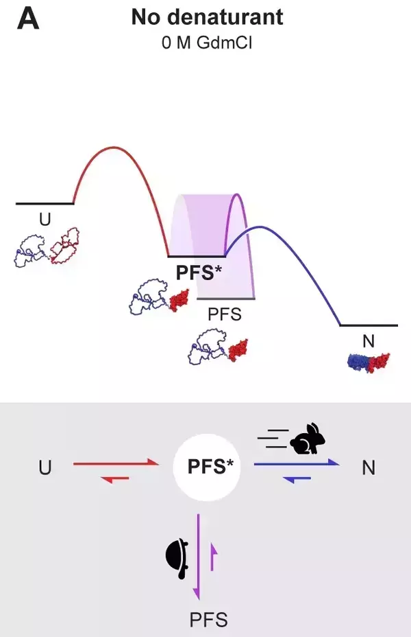
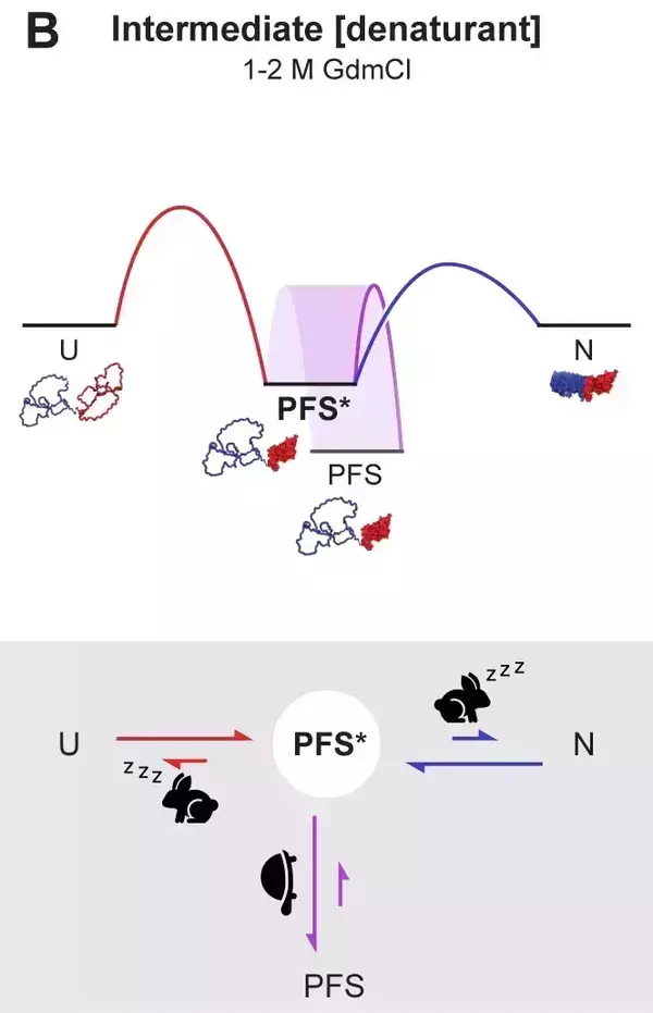
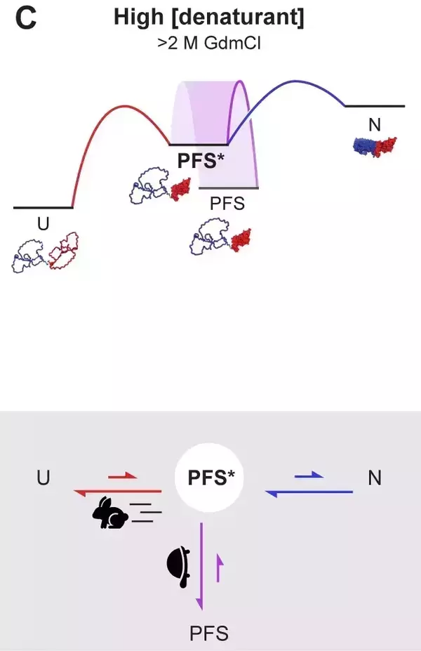
We are determining the folding and secretion mechanisms of autotransporters (ATs), the largest class of virulence proteins secreted from pathogenic Gram-negative bacteria. Gram-negative bacteria secrete ATs to their cell surface to mediate crucial pathogenesis functions, including biofilm formation, nutrient recruitment, host cell adhesion, and evasion of the host immune system. Our lab has shown that ATs initially adopt a disordered structure during their transport into the periplasm from N- to C-terminus, and fold to their native structure only after C- to N-terminal translocation across the bacterial outer membrane (OM). We have also shown that folding at the cell surface provides the driving force for a successful AT secretion across the bacterial OM (Drobnak et al. (2015) “Multiple driving forces required for efficient secretion of autotransporter virulence proteins”). However, other aspects of AT folding and secretion mechanisms remain unclear. For example, to what extent does an N-to-C versus C-to-N-terminal appearance vector prevents premature folding of ATs in the periplasm? More generally, how do AT structural features and folding properties promote efficient secretion? How do other aspects of the cellular environment contribution to successful AT secretion? We use biophysical approaches and biochemical assays to interrogate AT structural properties and test their impact on AT folding and secretion. We have developed new experimental approaches to structurally characterize intrinsically disordered proteins (IDPs) (Riback et al. (2017) “Innovative scattering analysis shows that hydrophobic disordered proteins are expanded in water”) and used these approaches to increase our understanding of how the dimensions of IDP conformational ensembles affect AT secretion (Baxa et al. (2024) “How hydrophobicity, side chains, and salt affect the dimensions of disordered proteins” and Bowman et al. (2020) “Properties of protein unfolded states suggest broad selection for expanded conformational ensembles”). Most recently, we have discovered that an AT protein can retain its C-to-N-terminal folding mechanism even in the absence of the spatiotemporal constraints of the cell (Luan & Clark (2024) “Discovery of an on-pathway protein folding intermediate illuminates the kinetic competition between folding and misfolding”). Collectively, insights from these projects have deepened our understanding of AT secretion mechanisms, opening avenues for novel strategies to combat diseases caused by Gram-negative bacterial infections.
Funding acknowledgement
Research progress in the Clark Lab is currently fueled by funding we receive from NIH, specifically the National Institute of General Medical Sciences. Patricia is a current recipient of an NIH Pioneer Award (DP1); she is the first and only Pioneer Award recipient in the state of Indiana since the program’s inception in 2004. Our past projects have also received funding from the National Science Foundation, the W.M. Keck Foundation, and the American Heart Association.